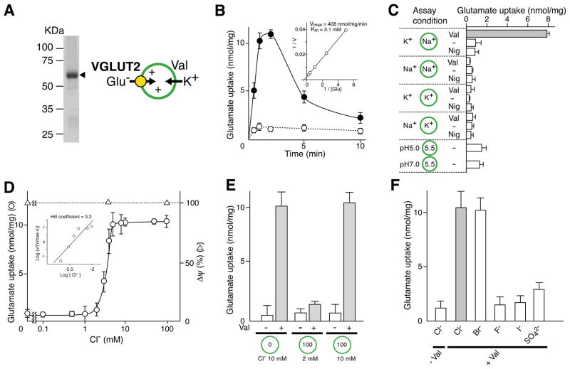
Figure 1. Cl− activates VGLUT2 externally.
A. Simple assay system for VGLUT. Proteoliposomes containing purified VGLUT2 (10 μg protein) were analyzed in an 11% polyacrylamide gel in the presence of SDS and visualized by Coomassie brilliant blue staining. The positions of molecular markers are indicated. The principle of Val-evoked glutamate uptake is also shown. B. Time course of glutamate uptake by reconstituted VGLUT2 was measured as described in Materials and Methods in the presence (closed circle) or absence (open circle) of Val. Kinetics of Val-evoked glutamate uptake at 1 min is shown in the inset. See also Figure S1. C. Energetics of glutamate uptake. Glutamate uptake was measured under the indicated ionic conditions. Nig, nigericin. D. Concentration dependence of Cl− on glutamate uptake. A sample was taken after 1 min. Δψ, measured as oxonol-V fluorescence quenching, is shown. The inset shows the Hill plot of glutamate uptake as a function of [Cl−]. E. Effect of internal and external Cl− on glutamate uptake. Proteoliposomes were prepared at the indicated concentrations of Cl− and suspended in buffer containing the indicated Cl− concentration. Val-evoked glutamate uptake was measured at 1 min. F. Cl− was replaced with the indicated anion and Val-evoked uptake of glutamate was measured at 1 min. Error bars represent mean ± SEM; n = 3.

