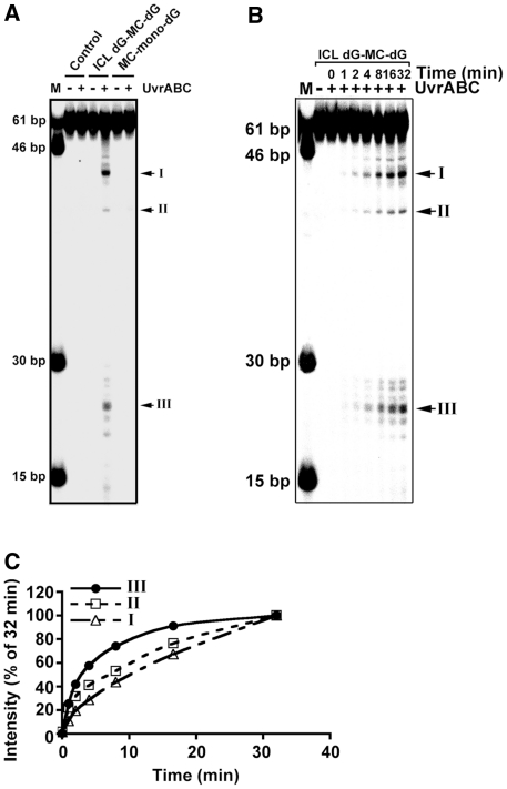
Figure 3.
Double-stranded DNA break formation resulted from UvrABC incision of an ICL dG-MC-dG adduct. (A) 32P-labeled substrate 1, substrate 2 and control 61-bp DNA fragments were reacted with UvrABC for 60 min and then separated in a nondenaturing polyacrylamide gel. (B) The kinetics of DSB formation resulted from UvrABC incision of an ICL dG-MC-dG DNA adduct. The 5′-end-32P-labeled 61-bp DNA fragments containing a site-specific ICL dG-MC-dG–DNA adduct were incubated with UvrABC nucleases as described in Figure 2. At different time periods of incubation, the resultant DNAs were separated in a nondenaturing polyacrylamide gel. The double-stranded DNA fragments standards are shown in lane M. (C) Quantitations. The band intensities at different times (It) shown in (B) were normalized to the intensity at 32 min (I32), which encompasses 90% of total activity. The data were plotted the same as in Figure 2.

