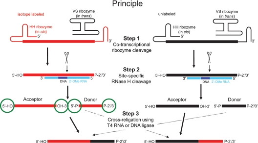
Figure 1.
Principle of the method comprising three reaction steps. The isotope labeled material is highlighted in red, the unlabeled material in black. In the 2′-O-methyl RNA/DNA chimera, the DNA is in dark blue and the 2′-O-methyl RNA in light blue. The termini of both acceptor and donor fragments are encircled in green. Scissors indicate RNase H cleavage sites. P-2′/3′ stands for a 2′/3′-cyclic phosphate.

