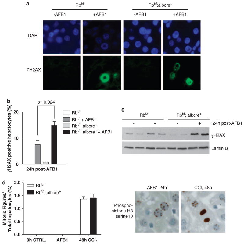
Figure 4.
Increased double stranded breaks (DSBs) and failure in mitotic progression in retinoblastoma (RB)-deficient livers exposed to aflatoxin B1 (AFB1). (a) Representative images of γ-H2AX were taken. (b) Plots depicting total positive hepatocytes for γ-H2AX. (c) Equal total protein was separated using sodium dodecyl sulfate polyacrylamide gel electrophoresis (SDS–PAGE), and the levels of γ-H2AX were detected. (d) Plot depicting mitotic figures present in AFB1-treated conditions, and carbon tetrachloride ligand 4 (CCl4) samples used as positive control. The images represent phospho-histone H3 serine10 stain.

