
Figure 1.
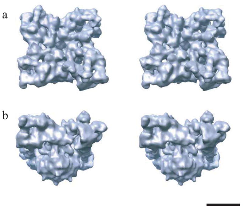
Stereo views of the 3D structure of RyR1 at 14 Å resolution. The structure is shown at an oblique angle as viewed from the cytoplasm (a) and in a side view (b) with the cytoplasmic side facing upward. The reconstruction was generated using ~22,000 individual particle images collected on a JEOL 1200EX electron cryomicroscope, equipped with a tungsten filament and operated at 100 kV under minimal dose conditions (5–7 e/Å2) and at the defocus range of 0.9–4.1 μm.3 The closed channel conformation was obtained by the depletion of Ca2+ with 1 mM EGTA.2,3 The threshold level corresponds to an enclosed protein mass of 2.5 MDa assuming a protein density of 1.35 g/cm3. The scale bar represents 100 Å.