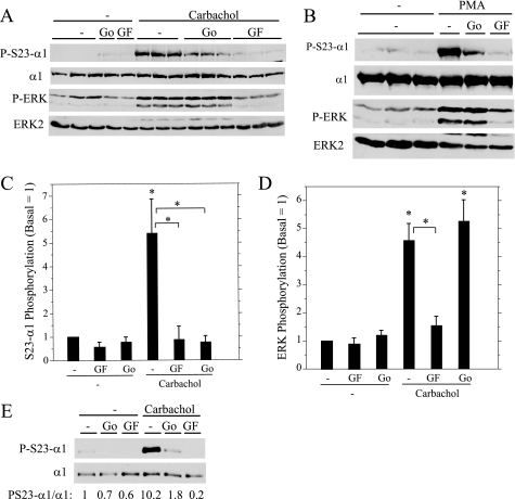
FIGURE 3.
Effect of PKC inhibitors on Ser23 α1 subunit (P-S23-α1) and ERK (P-ERK) phosphorylation. A and B, rat parotid acinar cells were treated for 10 min with vehicle (−), Go6976 (Go, 1 μm), and GF109203X (GF, 10 μm) followed by carbachol (10−5 m, 2 min) or PMA (100 nm, 2 min). Cell lysates were probed by Western blot as indicated. C and D, the phosphorylation of Ser23 α1 subunit (C) and ERK (D) in parotid acinar cells was quantified relative to basal conditions (no inhibitors). For Ser23 α1 (S23-α1) phosphorylation, n = 5–6, *, p < 0.05 versus basal or paired as indicated. For ERK phosphorylation, n = 5, *, p < 0.01 versus basal or paired as indicated. E, Par-C10 cells were treated with vehicle (−), Go6976 (1 μm, 20 min), and GF109203X (10 μm, 20 min) followed by carbachol (10−4 m, 5 min). α1 subunit immunoprecipitates were probed by Western blot as indicated and quantified relative to basal (no inhibitor).

