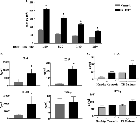
FIGURE 3.
Rv1917c-treated DCs stimulate CD4+ T cells to produce Th2 cytokines. A and B, DCs were cultured with GM-CSF and IL-4 alone (Control) or GM-CSF, IL-4, and 5 μg/ml of Rv1917c for 48 h. The Rv1917c-matured DCs were co-cultured with allogeneic CD4+ T cells at different DC to T cell ratios. After 4 days of co-culture, the cells were pulsed overnight with 0.5 μCi of [3H]thymidine to quantify T cell proliferation (A). Radioactive incorporation was expressed as counts/min (mean ± S.E. of quadruplet values). The data are representative for three independent donors. B, cell-free supernatants from DC:T cell co-cultures were analyzed for the cytokines IL-4, IL-5, IL-10, and IFN-γ. C, PBMCs obtained from tuberculosis patients (TB patients) (n = 11) and healthy control subjects (n = 4) were cultured with or without 5 μg/ml Rv1917c, and cell-free supernatants collected on day 4 were tested for concentrations of secreted IL-5 and IFN-γ. Data are represented as mean ± S.E. *, p < 0.05 versus control; **, p < 0.05 versus Rv1917c (Healthy Controls).

