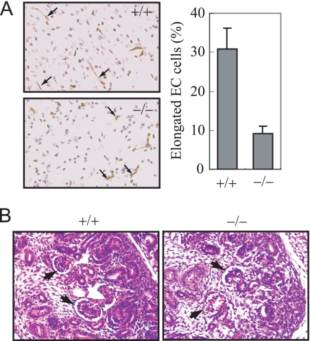
FIGURE 5.
Endothelial cell defects in Senp1−/− embryos. A, the brain sections from Senp1+/+ and Senp1−/− embryos at day 15 were stained with anti-CD31 to label endothelial cells (left panel). Right panel, elongated endothelial cell (EC) cells were counted and presented as the percentage of total CD31-positive cells in means ± S.D. of 9 sections from three pairs of littermates. B, H&E-stained sections of kidney from E15 Senp1+/+ (+/+) and Senp1−/− (−/−) embryo. Arrows indicate glomeruli.

