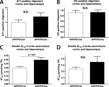
FIGURE 6.
Prefibrillar and fibrillar Aβ oligomers as well as soluble Aβ are not changed by the deletion of apoA-I in APP/PS1 mice. Soluble proteins were extracted from the cortices and hippocampi of APP/PS1/WT and APP/PS1/KO mice using TBS-based tissue homogenizing buffer as described under “Experimental Procedures.” A, prefibrillar Aβ oligomers were examined by dot blotting using A11 conformation-specific antibody. B, fibrillar Aβ oligomers were examined by dot blotting using OC conformation-specific antibody. In A and B, dot blot with 6E10 was performed and used to normalize the results of A11 and OC immunoreactivity. Shown is an ELISA for soluble Aβ40 (C) and Aβ42 (D). Note that Aβ40, unlike Aβ42, is significantly increased in APP/PS1/KO mice. Results are from 11–13 mice/group. Error bars, S.E.

