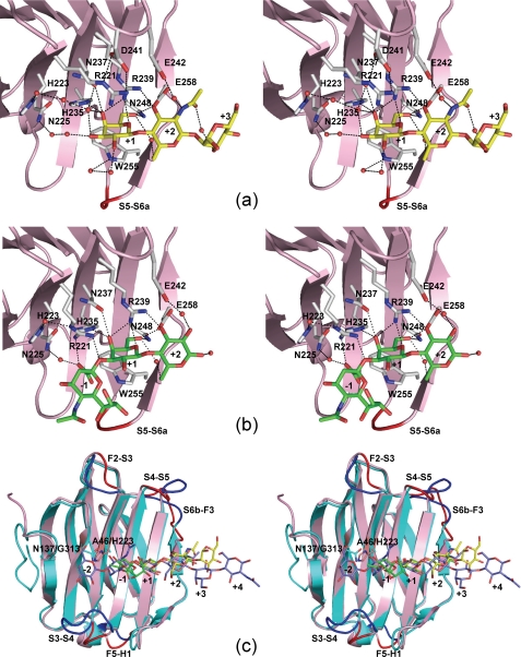
FIGURE 3.
Stereo views of the carbohydrate-binding sites of hG9C and a structural comparison between hG9C and hG9N. Selected hydrogen bonds with BIPA (a) and SiaLac (b) are shown by dotted lines. Although two conformers of Arg221 are found in hG9C-SiaLac, only conformer-2 is shown in b for clarity. c, superimposition of hG9C (pink) and hG9N (cyan) with the bound BIPA (yellow), SiaLac (green), and LN3 (blue). The loops with large deviations are indicated by dark colors.

