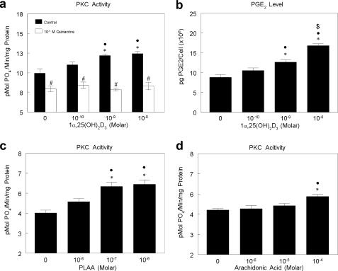
FIGURE 1.
Effect of 1α,25(OH) 2D3 on PGE2 production and PKC activity and role of PLA2 in PKC activation in MC3T3-E1 cells. a, MC3T3-E1 cells were treated with vehicle (ethanol) or 10−10, 10−9, or 10−8 m 1α,25(OH)2D3 with or without 10−5 m quinacrine (PLA2 inhibitor) for 9 min. PKC activity was normalized to total protein. b, MC3T3-E1 cells were treated with vehicle (ethanol) or 10−10, 10−9, or 10−8 m 1α,25(OH)2D3 for 30 min. Conditioned media were collected, and PGE2 was measured and normalized to cell number. c, MC3T3-E1 cells were treated with vehicle (ddH2O) or 10−8, 10−7, or 10−6 m PLAA for 9 min. PKC activity was normalized to total protein level. d, MC3T3-E1 cells were treated with vehicle (media) or 10−6, 10−5, or 10−4 m AA for 9 min. PKC activity was normalized to total protein level. *, p < 0.05, treatment versus control; ●, p < 0.05, 10−8 m and 10−9 m versus 10−10 m; $, p < 0.05, 10−8 m versus 10−9 m; #, p < 0.05 quinacrine versus control.

