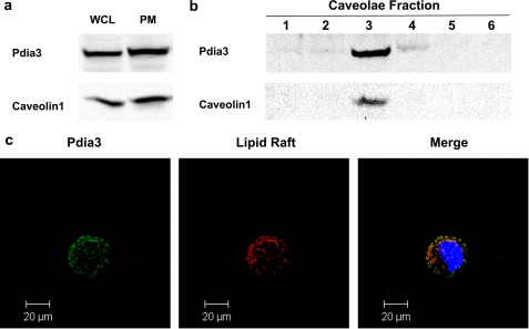
FIGURE 2.
Western blot and confocal microscope image of MC3T3-E1 cells. MC3T3-E1 cells were cultured as previously described. Whole cell lysates, plasma membrane fractions, and caveolae fractions were collected separately. Western blots against caveolin-1 and Pdia3 were performed. a, Western blot of whole cell lysates and membrane fractions. Thirteen fractions were collected; fractions one to six are shown. Caveolae exist in fraction three (b). c, confocal image of non-permeabilized MC3T3-E1 cells. Green: Pdia3; red: lipid rafts; yellow: merge of Pdia3 and lipid rafts.

