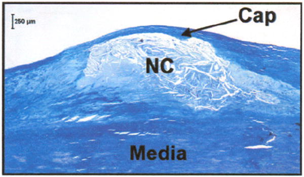
Fig. 1.

Histological section showing a necrotic core fibroatheroma (NCFA) with a thin fibrous cap (arrow) overlying a large necrotic lipid core (NC). The fibrous cap, predominantly composed of collagen and smooth muscle cells, is an important structural entity that determines the mechanical stability of the NCFA. Masson’s trichrome; original magnification 40×. Scale bar, 250 μm.
