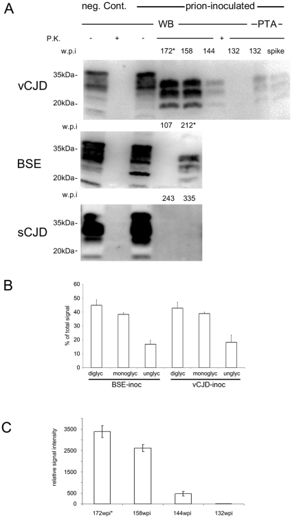
Figure 1. Biochemical analysis of PrPSc in the CNS.
A) Western Blot analysis for PrPSc (frontal cortex) of vCJD, BSE and sCJD infected animals. PrPSc could be demonstrated in the brains of preclinical vCJD and prion-diseased vCJD and BSE inoculated animals. In the vCJD cohort, PrPSc is detectable (using NaPTA) in subclinical state 40 weeks before onset of symptoms. sCJD inoculated macaques did not show PrPSc at any time point in cerebellum (data not shown) and frontal cortex in conventional as well as NaPTA enhanced Western blot. (* indicates prion-diseased animals). B) PrPSc-glycotype analysis demonstrates comparable glycotypes of vCJD and BSE when transmitted to primates. Densitometric measurement of relative band intensities for di-, mono- and unglycosylated form of PrPSc is shown in % of total signal. C) Quantification of PrPSc-signal shows initial exponential increase of PrPSc until 158 wpi when PrPSc levels off. Relative amounts of PrPSc are shown in arbitrary units as quantified in three independent experiments.

