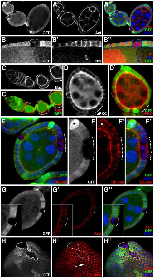
Figure 2. ph mutant cells within the follicular epithelium exhibit morphological and adhesion molecule modifications.
Analysis of mosaic ovarian follicles with ph0 mutant follicular cell clones integrated within the follicular epithelium (identified by the absence of GFP), using immunodetection to assay for distribution of cytoskeletal markers such as F-actin (Act) and Hu-li tai chao (Hts), apical markers such as Bazooka/Par3 (Baz) and aPKC, and adherens junctions components DE-cadherin (DE-cad) and Armadillo/β-catenin (Arm). All nuclei are stained with DAPI. (A–B) Apical accumulation of F-actin (A′) and lateral distribution of Hts (B′) are normal in the ph0 mutant clones (dotted lines). (C–D) The proteins Baz (C) and aPKC (D) are also correctly localized at the apical membrane in cells mutant for ph (dotted lines). (E) The distribution of the apical transmembrane protein DE-cadherin is affected in ph0 mutant clones (dotted lines) since significant amounts of basal accumulation are observed (F–F″, magnification of the clone in E). In F–F″, the asterisk indicates one of the polar cells which also exhibits some basal DE-Cadherin. (G–G″) Apical Armadillo/β-catenin distribution is not significantly disturbed in ph0 mutant cells (dotted lines). (H–H″) Adherens junction organization between ph wild-type epithelial cells produces a hexagonal honeycomb pattern of Armadillo/β-catenin staining in a surface view of a follicle (H′-arrow), while detection of this protein in clones of cells mutant for ph (H–H″ GFP negative, encircled) reveals cell morphological modifications including distinct elongation and apical constriction.

