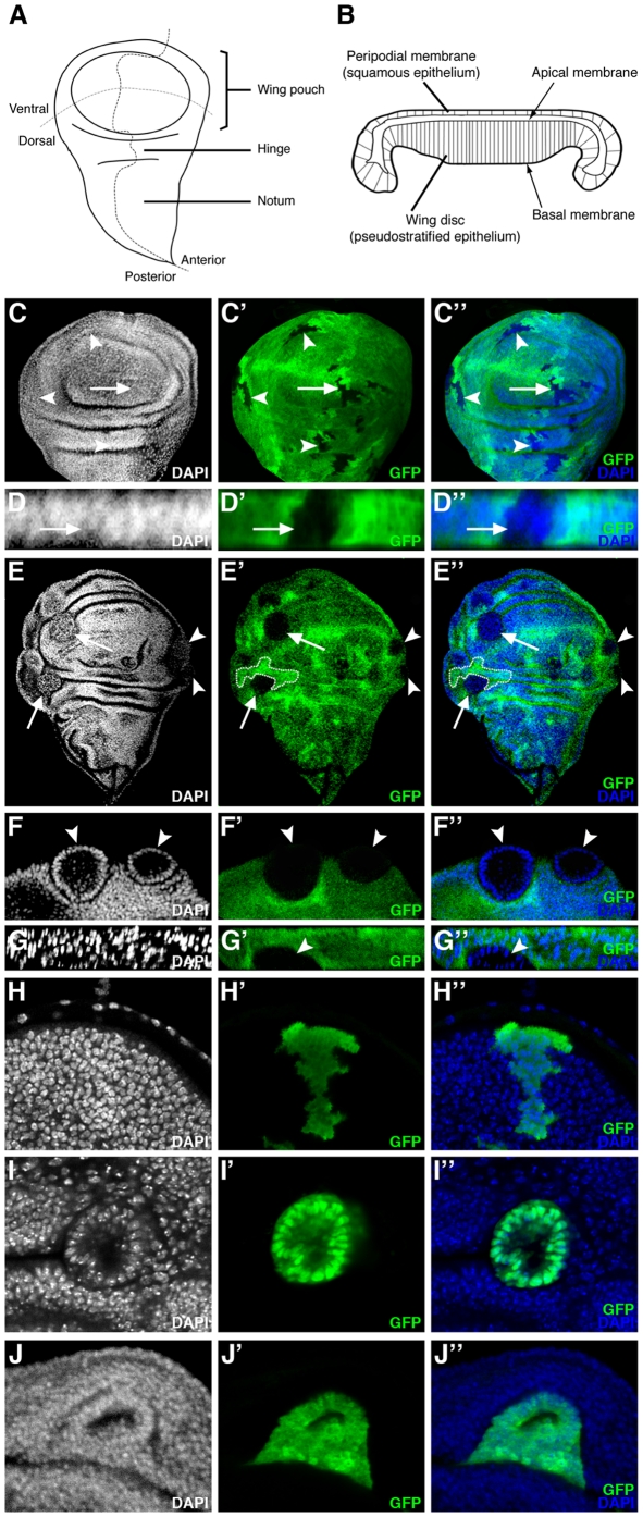
Figure 4. In the wing imaginal disc, ph misexpression clones sort-out from the wild-type epithelium.
(A) Drawing of a wing imaginal disc with compartments and presumptive territories indicated. (B) Drawing of a cross-section of a wing imaginal disc at the level of the wing pouch [47]. (C–G) Wing discs in which clones of cells have been induced which lack GFP (C–D) or lack both GFP and ph function (E–G). Panels C, E and F are XY confocal sections (orientation as in A). (C–C″) Wild-type clones lacking GFP form clones with wiggly borders evenly integrated in the epithelium (arrow and arrowheads). (D–D″) A XZ cross-section of the wing pouch (orientation as in B) of a wild-type clone (C–C″-arrow) showing GFP-negative cells integrated normally into the epithelial layer. (E–E″) ph0 homozygous clones lacking GFP segregate from surrounding control cells, disrupting the continuity of the epithelial layer and forming round clones with smooth borders (arrow and arrowheads). A twin clone with wiggly borders, indicated with dotted lines, is of approximately the same size as what is likely its corresponding ph0 clone. (F–F″) Magnified view of two mutant clones (arrowheads) from E (arrowheads), showing that ph0 cells are organized as monolayered spheres which bud off the wing disc. (G–G″) XZ cross-section (orientation as in B) of a ph0 clone (arrowhead) extruded basally as a cyst. (H,I,J) XY confocal views of wing discs in which clones of cells expressing GFP form wiggly borders with adjacent non-GFP expressing cells and are integrated normally in the disc epithelium (H), whereas clones expressing both GFP and the ph RNAi construct (I,) or GFP and the ph overexpression construct (J) form smooth borders with neighboring wild-type cells and a cyst structure. DAPI marks all nuclei.

