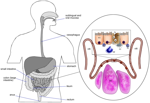
Figure 1. Schematic diagram of intestinal epithelium showing M cells, Peyer's patches, intestinal epithelial cells, and pathway of Ag transport.
DC, dendritic cells; IEC, intestinal epithelial cell (NU, nucleus); MC, M cell; IEL, intra epithelial lymphocytes; PP, Peyer's patches; MΦ, macrophages; Pv, particulate Ag in pinocytic vesicle of M cell.

