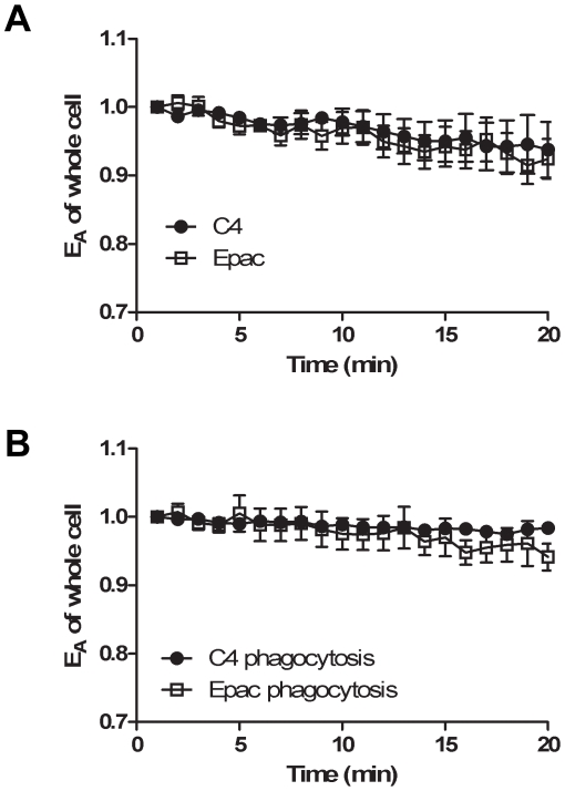
Figure 2. No change in total cAMP during phagocytosis.
RAW cells were transfected with plasmids for C4 control or the Epac-camps biosensor. Total cellular EA was measured over time and plotted relative to the first measured value. A. Measurements of unfed macrophages showed small decreases in FRET, indicating selective photobleaching of mCit. B. Transfected cells were fed opsonized targets and phagocytosis was synchronized as described in material and methods. No significant changes in cAMP were detectable during phagocytosis. Results are shown as mean ± SEM of 4–7 cells.

