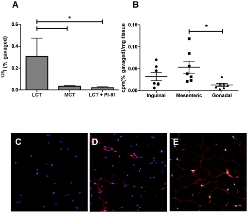
Figure 1. Intestinal absorption of dietary antigen (OVA) into adipose tissue.
(A) Fasted BALB/c mice (n = 4) were gavaged with 0.2 ml LCT, MCT, or LCT plus the inhibitor of chylomicron formation Pluronic L-81, and identical amounts of 125I-OVA. Gonadal adipose tissue was removed 60 minutes later and radioactivity was measured and normalized to tissue weight. (B) shows appearance of 125I-OVA into indicated adipose tissue samples 15 minutes after gavage of 125I-OVA in 0.2 ml 20% Intralipid. Asterisks indicate statistically significant differences (P<0.05) by one-way ANOVA with Bonferroni-adjusted post-hoc tests. (C–E) OVA immunostaining (red signal) in mesenteric adipose tissue of mice on OVA-free diets (C) or 1% OVA diets with low- (D) or high- (E) fat content. Blue signals represent DAPI-stained nuclei. OVA is mainly detectable in apparent endothelial cells, but staining can also be observed in other SVF cells and adipocytes.

