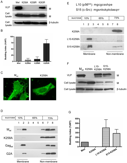
Figure 7. K258 is critical for NiV-M membrane association and budding.
(A) NiV-M K258 mutants are deficient in VLP budding. VLP and cell lysate samples were prepared from cells expressing wild-type M, K258A, K258R or K263R at 24 hpt as described in Materials and Methods . Immunoblotting was performed using an anti-FLAG monoclonal antibody, then the cell lysate blot was stripped and re-probed with an anti-β-tubulin antibody as loading control. Both K258A and K258R were expressed in the cells at similar levels compared to wild-type M, but they were absent from the VLPs. The experiment was repeated three times and representative results are shown. (B) Quantification of the budding index for the wild-type and mutant NiV-M proteins shown in (A). (C) Wild-type M localized to membrane patches and fine filopodia extensions while the K258A mutant did not. (D) K258A is deficient in membrane association. HEK293T cells expressing wild-type NiV-M, K258A, wild-type HIV Gag, or a myristoylation mutant of HIV Gag (G2A) were harvested at 24 hpt. Cell homogenates were loaded at the bottom of a 10–73% discontinuous sucrose gradient and ultracentrifuged for 16 hrs at 100,000×g. Eight fractions were collected from the top, and proteins were extracted using methanol/chloroform prior to immunoblotting with anti-NiV-M (in the case of Mwt and K258A) or anti-myc (Gagwt and G2A) antibodies. Membrane-associated proteins were collected at the interface between 10% and 65% sucrose as “fraction 2” as described previously [71]. (E) Fusion to L10 or S15, the membrane targeting N-terminal peptide sequence from p56lck and c-Src, respectively, restores membrane association to the K258A mutant. Membrane flotation centrifugation was performed as in (D). (F) Rescue of K258A budding by L10 and S15. VLP and cell lysate samples were prepared from HEK293T cells expressing the indicated constructs and examined by immunoblotting using a rabbit anti-NiV-M antibody. The cell lysate blot was also probed with an anti-β-tubulin antibody as loading control. The experiment was repeated three times. Representative blots are shown in (F), and the quantification of the budding indices is shown in (G).

