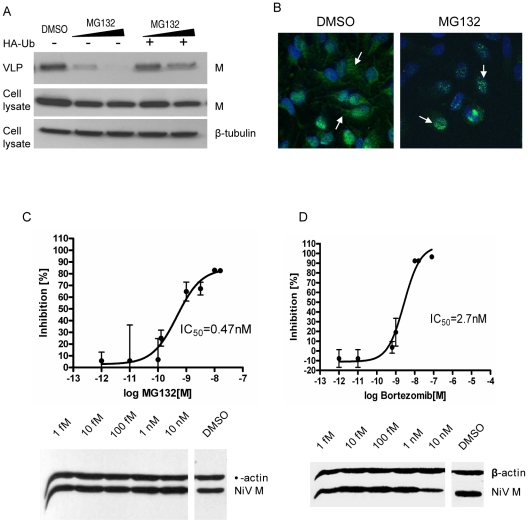
Figure 8. MG132 and bortezomib inhibit NiV-M nuclear export during live viral infection and reduce viral titers.
(A) NiV-M VLP budding in the presence of MG132. HEK293T cells expressing 3XFLAG-M (left three lanes) or 3XFLAG-M plus HA-Ub (right two lanes) were incubated with DMSO, 10 µM or 50 µM MG132 for 12 hrs, and VLPs produced during this period were harvested as described in Materials and Methods . VLPs and cell lysates were immunoblotted with an anti-FLAG antibody, then the cell lysate blot was stripped and re-probed with an anti-β-tubulin antibody as loading control. (B) MG132 altered M localization during live viral infection. HeLa cells infected with Nipah virus Malaysia strain were incubated with 50 µM MG132 or DMSO for 8 hrs starting from 15 hpi. Cells were then stained with an anti-M antibody and imaged on a confocal microscope. MG132 restricted M localization to the nuclear compartment. (C) and (D) Dose-response curves of Nipah viral titers in the presence of MG132 (C) or bortezomib (D). HeLa cells were incubated with NiV for 1 hr at 37°C and then fresh growth medium. 15 hpi, serial dilutions of MG132 or bortezomib were added, yielding final concentrations ranging from 10 nM to 1 fM. Considering the short half-life of bortezomib (9–15 hrs), it was re-added 12 hrs later. Supernatants were collected at 40 hpi and viral titers were determined by plaque assay. To calculate the 50% inhibitory concentration (IC50), the resulting data were fit to the sigmoidal dose-response curve (GraphPad Prism software version 4.00) using the equation: % inhibition = minimal inhibition + (maximal inhibition-minimal inhibition)/(1+10∧(LogIC50-Log drug concentration)). Results shown are from two independent experiments with triplicates for each data point. The infected cells were harvested, and the expression of cellular (β-actin) and viral (matrix) proteins was examined by immunoblotting.

