
Figure 2.
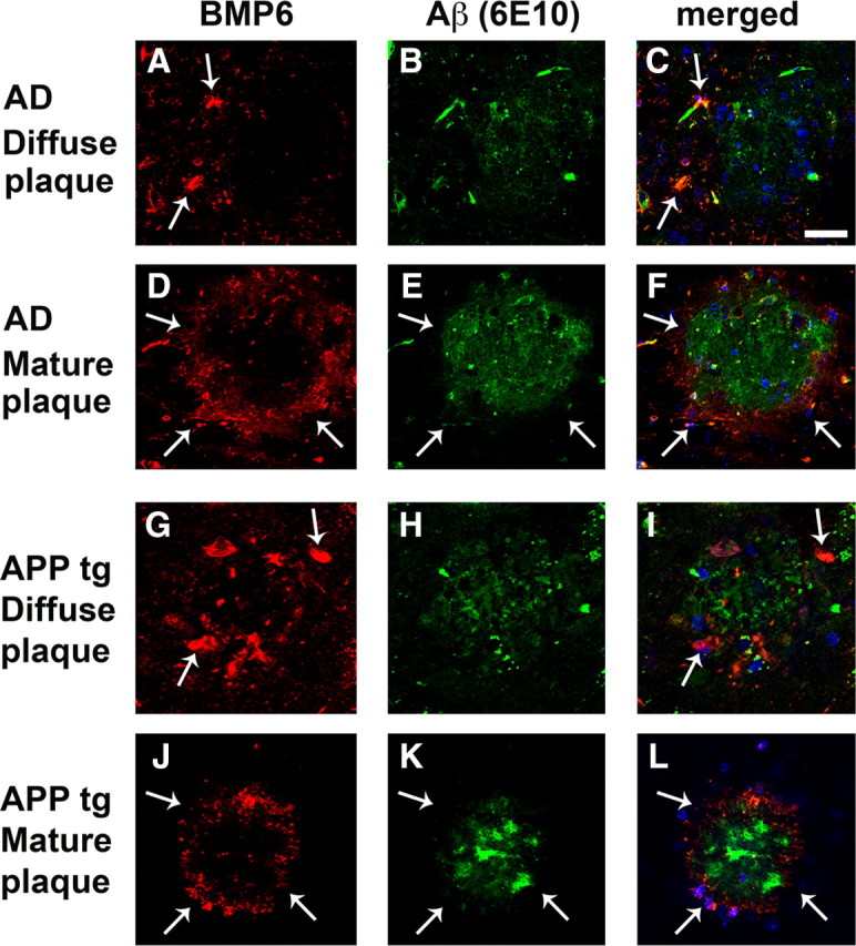
BMP6 immunoreactivity surrounds plaques in the hippocampus of AD patients and APP tg mice. Sections from the brains of severe AD cases or APP tg mice were immunolabeled with the monoclonal antibody against BMP6 (Millipore) detected with Tyramide Red and colabeled with an antibody against Aβ (6E10, Signet) detected with a FITC-tagged secondary antibody. All images are from the hippocampus (molecular and pyramidal cell layers), and sections were costained with DAPI (blue) to label cell nuclei. Scale bar, 100 μm for all images. A–F, Representative images showing BMP6 immunoreactivity (arrows, A, C, D, F) in a ring-like pattern surrounding Aβ-immunoreactive diffuse (A–C) and mature (D–F) plaques (arrows, E, F) in the brain of a human case with severe AD. G–L, Representative images showing BMP6 immunoreactivity (arrows, G, I, J, L) surrounding Aβ-immunoreactive diffuse (G–I) and mature (J–L) plaques (arrows, K, L) in the brain of an APP tg mouse.