
Figure 5.
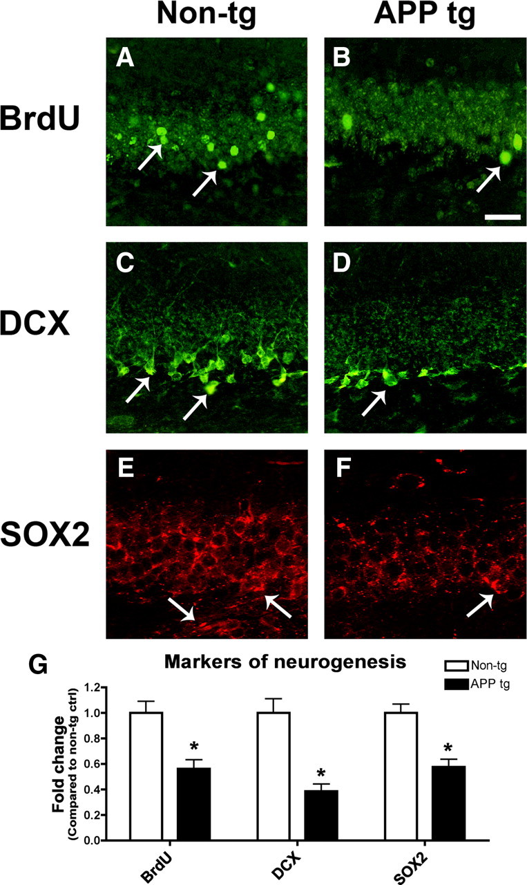
Markers of neurogenesis are reduced in the brains of APP tg mice. Sections from the brains of 6-month old non-tg control and APP tg mice treated with BrdU were immunolabeled with antibodies against BrdU or DCX detected with FITC-tagged secondary antibodies, or SOX2 detected with Tyramide Red and imaged by confocal microscopy. All images are from the hippocampal dentate gyrus. Scale bar, 30 μm for all images. A, B, Fewer BrdU-positive cells (arrows) were detected in the brains of APP tg mice compared to non-tg controls. C, D, Fewer DCX-positive cells (arrows) were detected in the brains of APP tg mice compared to non-tg controls. DCX-positive cells and processes were observed throughout the granular cell layer in non-tg control brains (C), but in APP tg mice DCX-positive cells and processes were less prominent, and were primarily observed only in the subgranular zone (D, arrow). E, F, Reduced SOX2 immunoreactivity in the dentate gyrus of APP tg mice compared to non-tg controls. SOX2 immunoreactivity was observed throughout the granular cell layer in the hippocampus of non-tg mice but to a lesser extent in APP tg mice (arrows). G, Semiquantitative analysis showing relative reductions in the numbers of BrdU-positive and DCX-positive cells, and decreased SOX2 immunoreactivity in the hippocampus of APP tg mice compared to non-tg controls (n = 4 animals per group, *p < 0.05 compared to non-tg controls by unpaired, two-tailed Student's t test).