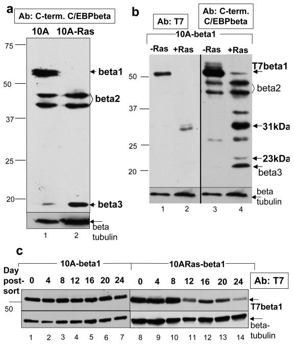
Figure 1.
Activated Ras(V12) negatively regulates C/EBPbeta1 expression in the immortalized MCF10A mammary epithelial cell line a. Immunoblot analysis of endogenous C/EBPbeta expression in MCF10A cells (lane 1) and MCF10A that express activated Ras(V12) (MCF10A-Ras) cells (lane 2). Equal amounts of total protein were loaded in each lane of a 12% sodium dodecyl sulfate – polyacrylamide gel electrophoresis (SDS-PAGE). The different isoforms of C/EBPbeta are indicated with arrows. Note - expression levels of the isoforms of C/EBPbeta in MCF10A cells can be somewhat variable depending on passage. Bars indicate the mobility’s of standard molecular weight markers, in kilo- Daltons (kDa), in all figures. Immunoblotting was performed with a C-terminal C/EBPbeta antibody (Santa Cruz C-19) at a dilution of 1:5000. The molecular weight markers used in this figure are the same as those used in previous papers which identify C/EBPbeta1 as a 55kDa protein. The remaining figures in this paper use a different molecular weight marker that shows C/EBPbeta1 having an apparent molecular weight of 52kDa. b. MCF10A and MCF10A-Ras cells infected with LZRS-T7-C/EBPbeta1-IRES-eGFP and sorted by FACS for GFP positive cells. C/EBPbeta1 is the only C/EBPbeta isoform produced by this retrovirus because the second ATG necessary to translate C/EBPbeta2 has been mutated along with a small open reading frame (ORF) required for translation of C/EBPbeta3 (Calkhoven et al., 2000). This immunoblot is three weeks after sorting. Equal amounts of total protein were loaded in each lane of a 10% SDS-PAGE. Immunoblot analysis of lanes 1 and 2 were performed with a T7 tag mouse monoclonal antibody (Novagen) at 1:10000 whereas lanes 3 and 4 are with an anti-C/EBPbeta C-terminal antibody (Santa Cruz C-19) at 1:5000. Arrows indicate full length p52-T7-C/EBPbeta1 along with smaller degradation products of T7-C/EBPbeta1. c. MCF10A (lanes 1-7) and MCF10A-Ras (lanes 8-14) cells infected with LZRS-T7-C/EBPbeta1-IRES-eGFP and sorted by FACS for GFP positive cells. Cell lysates were prepared every four days for 24 days. Equal amounts of total protein were loaded in each lane of a 10% SDS-PAGE. Immunoblot analysis was performed with the T7-tag antibody (Novagen) at a dilution of 1:10000. Arrows indicate full length p52-T7-C/EBPbeta. Immunoblot analysis with the beta-tubulin antibody is shown as a loading control (Sigma T7816 at 1:10000) (beta = C/EBPbeta, 10A = MCF10A)

