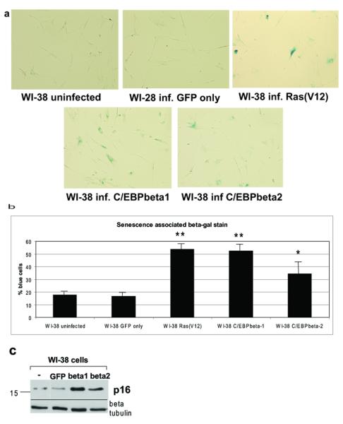
Figure 6.
C/EBPbeta1, and to a lesser extent C/EBPbeta2, induce senescence in WI-38 human fibroblasts a. WI-38 cells were infected with LZRS-GFP, LZRS-T7-C/EBPbeta1-IRES-eGFP, LZRS-T7-C/EBPbeta2-IRES-eGFP, or pBABE Ras(V12)-puromycin. Six days post-infection 60% confluent cells were fixed in 60mm plates and stained for with the senescence associated beta-galactosidase kit as per manufacturers instructions (Cell Signaling Technology). Five fields per plate were imaged with a light microscope with representative photomicrographs displayed. b. Quantitative comparison of senescence associated beta-galactosidase stain. This experiment was repeated three times with standard deviation represented by error bars. Quantitative comparison using the student t test indicates that there is a statistically significant difference in blue staining between the Ras(V12), T7-C/EBPbeta1, and T7-C/EBPbeta2 expressing WI-38 cells versus uninfected. ** p < 0.006, * p < 0.03. c. WI-38 cells were infected with LZRS-GFP, LZRS-T7-C/EBPbeta1-IRES-eGFP, or LZRS-T7-C/EBPbeta2-IRES-eGFP. Six days post-infection cell lysates were made and run on a 10% SDS-PAGE. Equal amounts of protein were loaded in each lane. Immunoblot analysis was carried out with an anti-p16INK4A antibody (Santa Cruz). Immunoblot analysis for beta-tubulin was performed as a loading control (Sigma T7816).

