Figure 5.
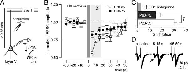
(A) Recording arrangement designed to study depolarization-induced suppression of excitation (DSE) in deep-layer pyramidal neurons of the medial prefrontal cortex. DSE was assessed by measuring the effect of transient (5 s) postsynaptic depolarization (+10 mV) on electrically-evoked EPSCs elicited by local stimulation of excitatory inputs onto the apical dendrite. (B) Time course of evoked-EPSC amplitude recorded in pyramidal cells from juveniles (P28–35; n=7 cells/5 rats) and adults (P60–75; n=8 cells/6 rats) before, during and after DSE. The results show that the magnitude of DSE is more robust in the juvenile prefrontal cortex (B'). (C) Bar graph depicting the averaged amplitude inhibition in the first three measurements during DSE as shown in B' and the results of the CB1 receptor antagonist treatment. Bath application of the CB1 receptor antagonist AM-251 (4 μM) blocked the DSE in all cells tested (n=8 cells/8 rats). Group effect (P28–35 vs. P60–75, P28–35 vs. AM-251 and P60–75 vs. AM-251): **P<0.01. (D) Electrophysiological traces of evoked-EPSCs recorded before (baseline), during (5–15 s) and after (45–50 s) DSE. Note the reduction of the first EPSC amplitude (arrow) while the second EPSC remained unchanged.

