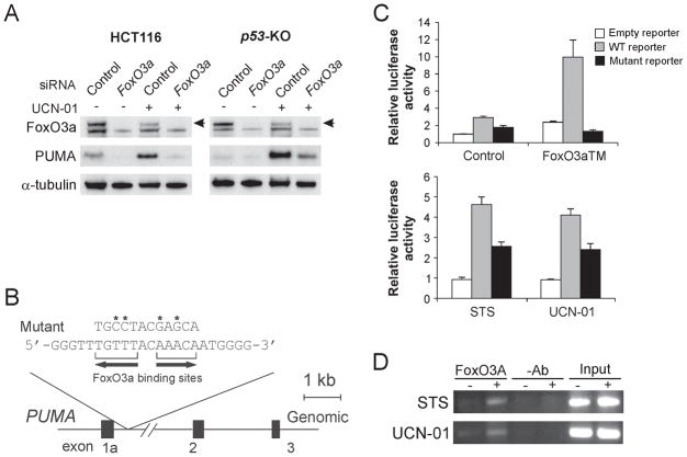
Figure 2. FoxO3a directly activates PUMA transcription following UCN-01 treatment.
A. p53-KO HCT116 cells were transfected with either a control scrambled siRNA or a FoxO3a siRNA for 24 hr, and then treated with 1 μM UCN-01 for 24 hr. FoxO3a and PUMA expression were probed by Western blotting. Arrow denotes FoxO3a-specific band. B. Schematic representation of the genomic structure of PUMA highlighting the two FoxO3a binding sites (AAACA) within the first intron. Asterisks indicate binding site mutations. C. Upper: p53-KO HCT116 cells were transfected with a reporter plasmid containing the WT or mutant FoxO3a binding sites in the PUMA promoter, along with a FoxO3a triple mutant (FoxO3aTM) expression construct or the control empty vector. The reporter activities were measured by luciferase assay on the next day. Lower: p53-KO HCT116 cells were transfected overnight with the reporter containing the WT or mutant FoxO3a binding sites, and then treated with 60 nM staurosporine (STS) or 0.5 μM UCN-01 for 16 hr. Luciferase activity was quantified. D. Chromatin immunoprecipitation (ChIP) was performed on fixed p53-KO HCT116 cells following 8 hr 60 nM STS or 1 μM UCN-01 treatment. An antibody specific for FoxO3a or no antibody was used to show specificity. PCR was carried out using primers surrounding the FoxO3a binding sites in the PUMA promoter.

