
Fig. 3.
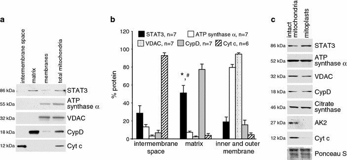
STAT3 is present in the mitochondrial matrix. a Rat LV SSM were subfractionated into the proteins of the intermembrane space, the matrix, and the inner and outer membrane (membranes). Western blot analysis was performed for STAT3 and marker proteins of different submitochondrial compartments (intermembrane space: Cyt c, matrix: CypD, outer membrane: VDAC, inner membrane: ATP synthase α). Total mitochondrial protein extracts were used as control. bBar graphs representing the percentage of protein in mitochondrial compartments. The total amount of pixels from each subfractionation protocol was set as 100%. *,#p < 0.05 for STAT3 immunoreactivity in the matrix versus the intermembrane space and the inner and outer membrane, respectively. c Rat LV SSM were incubated in hypo-osmotic buffer to induce mitochondrial swelling and subsequently characterized by western blot analysis. Swelling induces loss of marker proteins for the intermembrane space (adenylate kinase 2, AK2, and cytochrome C, Cyt c), whereas immunoreactivities for membrane proteins (ATP synthase α, inner membrane) or VDAC (outer membrane), matrix proteins [citrate synthase, cyclophilin D (CypD)] as well as STAT3 immunoreactivity were unchanged