
Fig. 9.
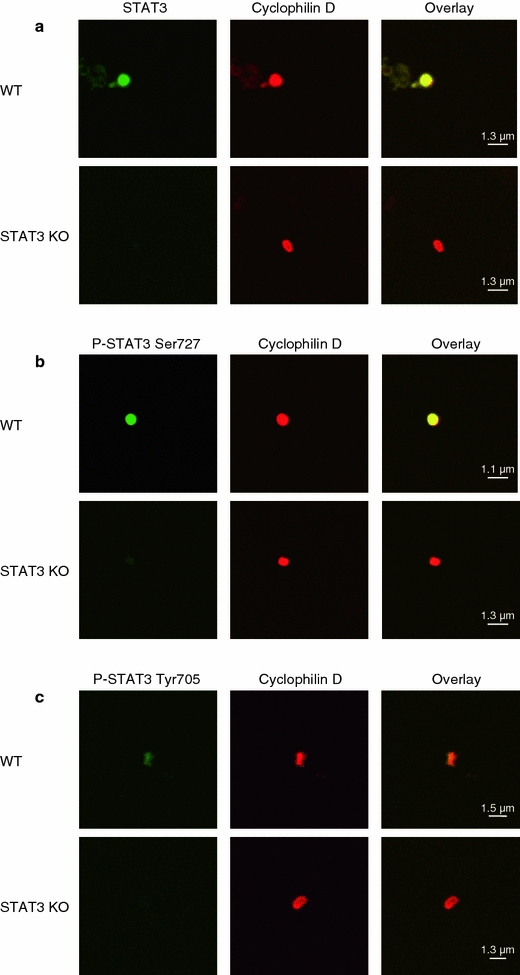
Total and phosphorylated STAT3 are present in mouse left ventricular (LV) mitochondria. a LV subsarcolemmal mitochondria (SSM) of WT (n = 6) and STAT3-KO mice (n = 8) were stained with antibodies against STAT3 (green) and the mitochondrial marker cyclophilin D (red) and were analyzed by confocal laser scan microscopy. Overlay pixels are shown in yellow. Whereas in SSM isolated from the LV of WT mice about 91% (347 mitochondria counted) of the ATP synthase positive mitochondria were also positive for STAT3, only 13% (248 mitochondria counted) of the SSM had immunoreactivity for STAT3 in STAT3-KO mice. b SSM from the LV of WT (n = 4) and STAT3-KO mice (n = 4) were stained with antibodies against STAT3 phosphorylated at Ser727 (green) and the mitochondrial marker cyclophilin D (red) and were analyzed by confocal laser scan microscopy. Overlay pixels are shown in yellow. c SSM from the LV of WT (n = 4) and STAT3-KO mice (n = 4) were stained with antibodies against STAT3 phosphorylated at Tyr705 (green) and the mitochondrial marker cyclophilin D (red) and were analyzed by confocal laser scan microscopy. Overlay pixels are shown in yellow