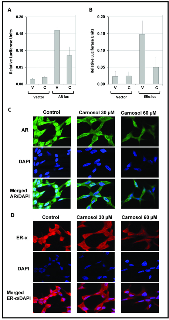
Figure 4.
LNCaP cells were transiently transfected with an ~6-kb AR promoter-luciferase reporter plasmid in a pGL3 Basic Vector or ~5.8-kb ER-α promoter-luciferase reporter plasmid in a pGL2 Basic Vector. 18 h after transfection, cells were treated with DMSO (vehicle) or 30 µM carnosol and analyzed for Luciferase activity after 24 h treatment with carnosol. The mean represents three individual samples with bars representing standard deviation (SD); * = p <0.01. C–D, LNCaP cells were grown in chamber slides at 100,000 cells per chamber overnight and treated with carnosol (30 µM) for 24 h. For labeling, anti-AR antibody (Santa Cruz Biotechnology, Santa Cruz, CA.) and Alexa Fluor 488 goat anti-rabbit IgG (Invitrogen) were used as primary and secondary antibodies after 24 h carnosol treatment, respectively. For labeling anti-ER-α (Cell Signaling Technology, Danvers, MA) and Alexa Fluor 594 goat anti-mouse IgG were utilized after 24 h treatment. 4,6-Diamidino-2-phenylindole (DAPI) was used to counter stain the nucleus.

