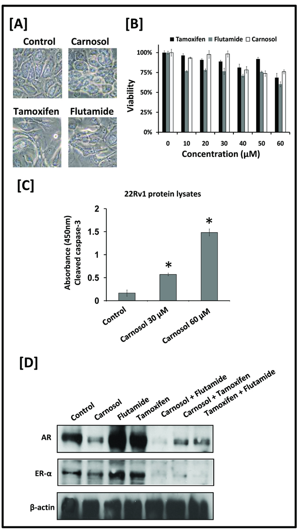
Figure 5.
A, Prostate epithelial cells (PrEC) were treated with DMSO (i.e. vehicle control), carnosol (30 µM), flutamide (30 µM), or tamoxifen (30 µM). Phase contrast images at 40× magnification were taken 48 h after treatment. B, PrEC cells were treated with increasing concentrations of carnosol, tamoxifen, or flutamide and MTT assay was performed to determine the effect on cell viability. Mean represents three individual samples; bars represent standard deviation. C, Cleaved (activated) caspase-3 was detected by ELISA. 22Rv1 cells were treated with vehicle, carnosol 30 uM, Carnosol 60 uM for 24 hours and protocol was followed per manufacturer’s directions. Mean represents average of three individual samples; bars represent standard deviation. *, P <0.01 of carnosol treated samples versus control. D, LNCaP cells were grown to 60–70% confluence and medium was replaced containing carnosol, flutamide, and tamoxifen, or combinations of carnosol/flutamide, carnosol/tamoxifen, or flutamide/tamoxifen. Total cell lysates were prepared and 40 µg of protein were subjected to SDS-PAGE followed by Western blot analysis. Equal loading of protein was confirmed by stripping the immunoblot and reprobing it for β-actin.

