
Figure 2.
Histologic Analysis of Distal Femur Cartilage from Case R00-394 and Control
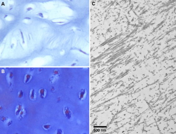
(A and B) Light microscopy. Cartilage was decalcified, embedded in paraffin, and stained with von Kossa trichrome. The fibrochondrogenesis case (A, 32 weeks gestation) exhibits fibroblastic chondrocytes and a fibrous intercellular matrix not present in the 28-weeks-gestation control (B). Magnification is 40×.
(C) Transmission electron microscopy showing frayed and irregular collagen fibrils.