
Figure 4.
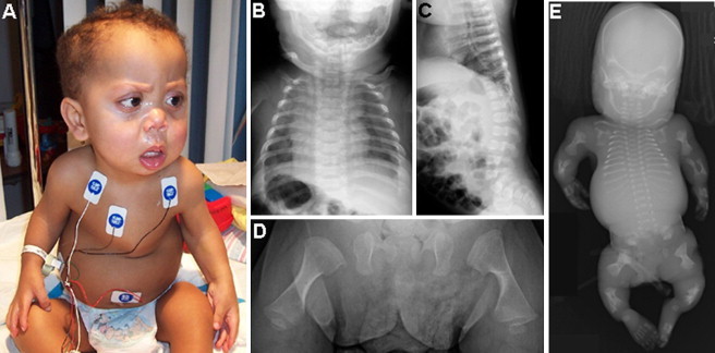
Clinical Photograph and Radiographs of Fibrochondrogenesis Cases from Family R06-573
(A) The surviving boy (R06-573C) at 19 months of age, presenting with a flat midface, small nose with anteverted nares, and a small bell-shaped thorax.
(B) An anterior-posterior chest radiograph of the boy at 2 months of age shows short and wide ribs with metaphyseal cupping.
(C) A lateral-view radiograph of the spine shows flat vertebral bodies.
(D) An anterior-posterior radiograph of the femurs shows short long bones with broad metaphyses.
(E) Anterior-posterior radiograph of the first affected fetus (R06-573A) in the family at 24 weeks gestation.