
Figure 7.
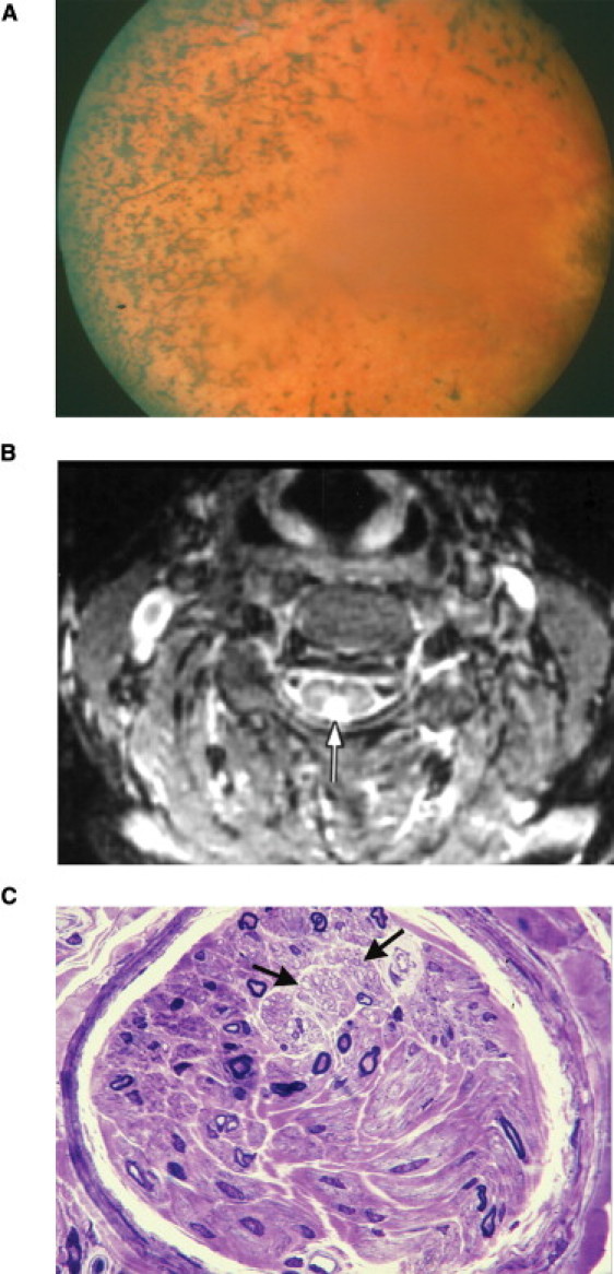
Phenotypic Features of Individuals with PCARP and FLVCR1 Mutations
(A) Fundus photograph of individual 30 from the American family (Figure 1) at age 39, showing typical bone-spicule pigmentation and retinal vessel attenuation seen in RP.
(B) Axial MRI scan (TR-3000, TE-30, T1-150) with a white arrow pointing toward an abnormal hyperintense signal in the posterior aspect of the spinal cord of individual 30 (Figure 1) at age 39.
(C) A semithin cross-section of a sural nerve fascicle showing severe loss of large myelinated fibers in individual IV-14 (Figure 2) at age 35. The density of myelinated fibers was 850 per mm2, with a complete loss of myelinated fibers having diameters larger than 8 μm. The two black arrows point to an area with preserved unmyelinated fibers (toluidine blue, 63× objective). Scale bar represents 10 μm.