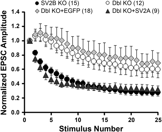
Fig. 1.
Acute expression of synaptic vesicle protein 2A (SV2A) restores normal synaptic depression in hippocampal neurons from SV2A/B double (Dbl) knockout (KO) mice. Hippocampal neurons from SV2A/B double KO mice were infected with Semliki Forest virus containing cDNA encoding SV2A + enhanced green fluorescent protein (EGFP) (solid triangles), or EGFP alone (shaded diamonds). Synaptic responses to depolarizing trains were assayed within 18 h of infection. Shown are average excitatory postsynaptic current (EPSC) amplitudes in response to a 10-Hz train of depolarizing pulses, normalized to the amplitude of the first response. Values are means ± SE. Neurons expressing SV2A displayed robust synaptic depression, whereas those expressing EGFP alone did not. Also shown for comparison are responses from uninfected SV2B KO, which have a wild-type (WT) phenotype (solid circles) and SV2A/B double KO neurons (open circles).

