Fig. 1.
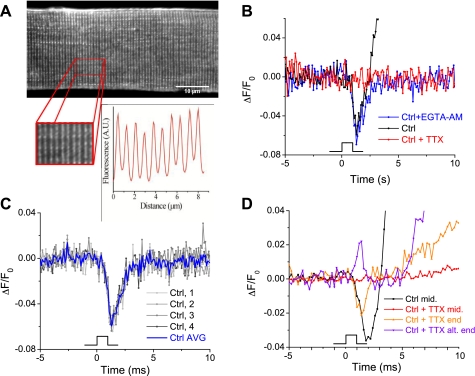
Optical recordings of the propagated action potential in flexor digitorum brevis (FDB) fibers. A: FDB fiber stained with di-8-aminonaphthylethenylpyridinium (di-8-ANEPPS) demonstrates lipophilic staining of the sarcolemma and T-system. Inset shows the doublet staining pattern with matched fluorescent intensity profile characteristic of the T-tubules. B: ΔF/F0 recordings of di-8-ANEPPS-stained fibers excited with a 1-ms field stimulus to generate an action potential (AP). Under control conditions, fibers demonstrated a large movement artifact during and after repolarization (black trace). This was abrogated by loading fibers with 50 μM EGTA-AM to buffer released calcium and prevent contraction (blue trace). Treatment of fibers with 100 nM TTX eliminated any fluorescence change (red trace). C: with the use of EGTA-AM/di-8-ANEPPS coloading protocol, AP recordings were highly reproducible and 4 APs were averaged for each trial to improve signal to noise. D: electrical stimulus artifacts in FDB fibers. Electrical signals elicited in the longitudinal middle of the FDB fiber always showed a downward deflection regardless of stimulus polarity (black trace) and were completely abrogated by TTX (red trace). However, even in the presence of TTX, electrical artifacts were seen at the ends of many fibers (orange and purple trace). These local signals alternated directionality with alternating stimulus polarity and promoted local depolarization, which triggered Ca2+ release and local contraction [evidenced by the smaller movement artifacts following stimulation compared with control conditions, which elicited a homogenous fiber contraction (black trace)].

