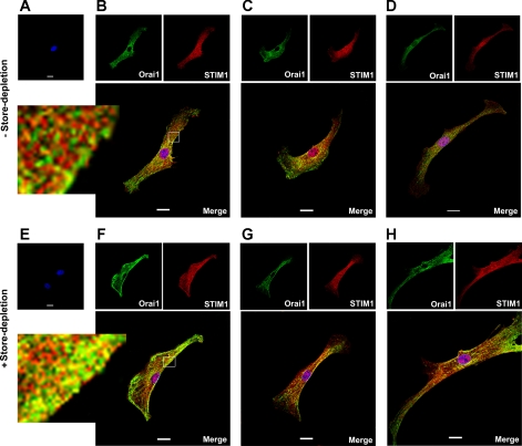
Fig. 6.
Colocalization of Orai1 and STIM1 in mouse PASMCs. A–D: staining of mouse cultured PASMCs after exposure of live cells with normal bath physiological salt solution (PSS). A: omission of Orai1 and STIM1 antibody resulted in no Orai1 or STIM1 staining. B–D: three representative cells dual-labeled with anti-Orai1 antibody (green) and STIM1 antibody (red). Orai1 and STIM1 colocalization (yellow/orange) is shown in the merged images. E–H: staining of mouse cultured PASMCs after exposure of live cells with Ca2+-free PSS containing 10 μM CPA. E: omission of Orai1 and STIM1 antibody resulted in no Orai1 or STIM1 staining. F–H: three representative cells dual-labeled with anti-Orai1 antibody (green) and STIM1 antibody (red). Colocalization of Orai1 and STIM1 is more apparent (yellow/orange) after store-depletion as shown in the merged images. Nuclei were stained with DAPI (blue). Experiments were performed in 3 separate immunostaining procedure, each with duplicate coverslips. Scale bars, 20 μm.

