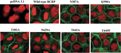
Fig. 3.
Confocal microscopy of HEK-293 cells stably expressing wild-type and mutant BCRP. The cellular localization of wild-type and mutant BCRP in HEK-293 cells (shown in green) was determined by immunofluorescence detection using the BCRP-specific antibody BXP-21, as described. Cell nuclei were stained with DAPI and are shown in red. No green fluorescence was detected in the vector control cells. Representative areas of HEK-293 cells expressing wild-type BCRP and the mutants N387A, Q398A, T402A, N629A, T642A, and Y645F are shown. Images have been enhanced for maximal contrast between the black background and green fluorescence and were not intended for quantitative determination of BCRP expression.

