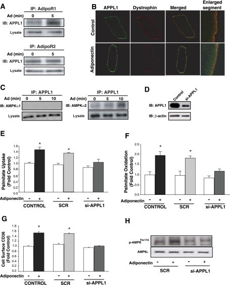
Fig. 2.
Functional participation of APPL1 in Ad's physiological actions in isolated rat cardiomyocytes. Representative immunoblots (IB) showing interaction between APPL1 and adiponectin receptor 1 (AdipoR1) or AdipoR2 (A) and AMPKα1 or AMPKα2 catalytic subunits (C). B: representative confocal images (optical single slice) showing individual and merged staining of cell surface APPL1 (green) or sarcolemma marker dystrophin (red). Enlarged segment is an optical single slice, magnification ×60. D: immunoblot showing knockdown of endogenous APPL1 (si-APPL1) compared with nontransfected (Control) neonatal rat cardiomyocytes. Palmitate uptake (E), palmitate oxidation (F), and CD36 translocation (G) in isolated neonatal rat cardiomyocytes not transfected (Control) or transfected with an unrelated siRNA (SCR) or siRNA against APPL1 (si-APPL1) and were treated with or without 10 μg Ad for 1 h (E and F) or 30 min (G). H: representative immunoblot showing AMPK phosphorylation (Thr172) in isolated neonatal rat cardiomyocytes transfected with SCR or si-APPL1 and subsequently treated with or without 10 μg Ad for 10 min. Data are means ± SE of ≥4 independent experiments and are expressed relative to Control (0 min). *P < 0.05 vs. Control (0 min).

