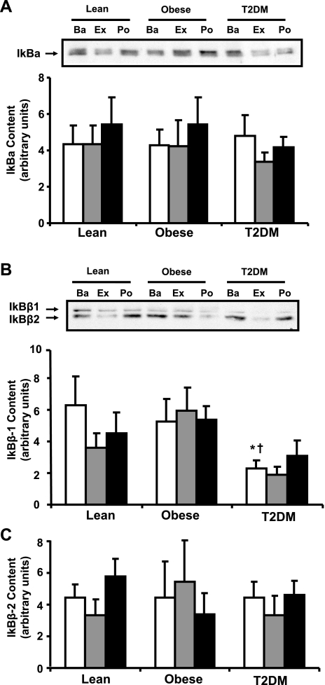
Fig. 4.
IκB protein content in muscle. Biopsies were done at Ba, Ex, and Po, and IκBα (A), IκBβ1 (B), and IκBβ2 content (C) were measured by Western blotting. Graphic data are means ± SE. Representative blots are shown for 1 subject/group; *P < 0.05 vs. lean at baseline; †P < 0.05 vs. obese at baseline.

