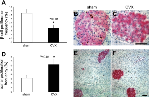
Fig. 3.
β-Cell proliferation is reduced in CVX rats. A: 1 wk following the CVX surgery, the β-cell proliferation frequency was reduced to 50% of the sham values, as determined by Ki-67 and insulin immunostaining. Age-matched, untouched control rats exhibited β-cell proliferation values identical to the sham controls (data not shown). B: representative fields of a sham control rat islet. C: CVX rat islet demonstrating proliferating cells. Scale bar, 20 μm. D: acinar cell proliferation increased 2-fold in the CVX group. E: representative low-power fields of a sham control pancreas. F: CVX rat pancreas showing increased Ki-67 staining in acinar cells of the latter. Scale bar, 20 μm. Brown, Ki-67 immunoreactivity; magenta, insulin; blue, nuclei.

