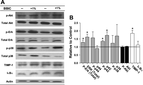
Fig. 5.
Changes in MAPKs, Akt, and TIMP-1 in mdx mice given control or 1% BBIC containing diet. A: representative immunoblots of activity and expression. p-Akt, phospho-Akt; p-Erk, phospho-ERK; p-p38, phospho-p38. B: relative change graph of MAPKs, Akt, and TIMP-1 to show growth signaling changes and endogenous regulation to suppress increased metalloproteinase activity in mdx (n = 4). *P < 0.05 vs. control-fed mdx.

