
Fig. 8.
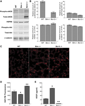
Increased eNOS expression in the absence of bcl-2. Protein lysates (20 μg) from wild-type, bim−/−, and bcl-2−/− lung EC were analyzed by Western blot analysis for expression of phospho-eNOS, total eNOS, HSP90, phospho-Akt1, and total Akt1. β-catenin expression was assessed as a loading control (A). Quantitation of these data are shown in B. C: fluorescence photomicrographs of lung P28 wild-type, bim−/−, and bcl-2−/− mice stained with anti-eNOS. Note the increased eNOS staining in lung from bcl-2−/− mice. In D, the intracellular NO production was determined as analyzed by DAF-FM fluorescence. In E, an immunoassay was used to determine VEGF levels (pg/ml) in lung EC from wild-type, bim−/−, and bcl-2−/− mice. Experiments were repeated twice with 2 different isolations of lung EC cells with similar results. Note increased levels of total eNOS and NO levels in bcl-2−/− lung EC and increased VEGF levels in bim−/− lung EC. *P, **P < 0.05.