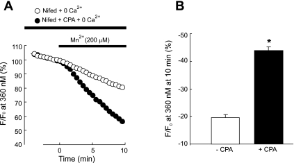
Fig. 3.
A: time course of fura-2 fluorescence at 360 nm normalized to values at time 0 before and after administration of MnCl2 (200 μM) to distal PVSMC perfused with Ca2+-free KRB solution containing 10 μM CPA, 1 mM EGTA, and 5 μM nifedipine (+CPA; n = 7 experiments in 177 cells) and in control cells, which did not receive CPA but were otherwise treated similarly (−CPA; n = 5 experiments in 137 cells). B: average decrease in fura-2 fluorescence at 10 min after administration of MnCl2 to cells shown in A. *P < 0.0001 vs. cells absent of CPA.

