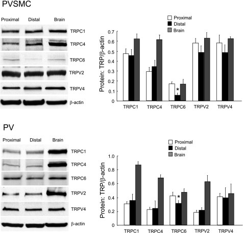
Fig. 9.
Expression of TRPC and TRPV proteins in rat proximal and distal PVSMC and PV as determined by Western blotting using rat brain as positive control. Data show representative blots for TRPC1, TRPC4, TRPC6, TRPV2, TRPV4, and β-actin in rat distal PVSMC (top: n = 5) and PV (bottom: n = 3). *P < 0.05 vs. respective proximal cells or tissue.

