Fig. 7.
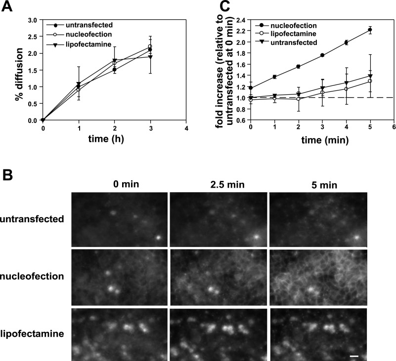
Tight junction fence, but not gate function, is disrupted in nucleofected cells. A: kinetics of transepithelial [3H]inulin diffusion across filter-grown MDCK cells. B: FM4–64FX (100 μM) was added to the apical chamber of filter-grown MDCK cells that had been previously treated as indicated. Cells were imaged every 5 s for 5 min and optical slices collected 2.5 μm above the level of the filters at 0, 2.5, and 5 min after addition of the dye are shown. All images were acquired and processed using identical conditions. The bright spots represent out-of-focus fluorescence from apoptotic cells above the cell monolayer and were especially prominent in lipofectamine-treated samples. Bar = 10 μm. C: change in intensity of FM4–64FX staining at the lateral surface over time was quantified in 2 independent experiments and is plotted relative to the initial intensity measured at time 0 in untransfected cells.

