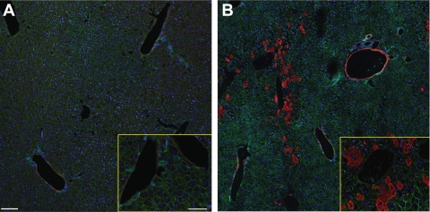
Fig. 2.
Complement factor 3 deposition in liver after HS/T. Immunofluorescence staining showed intense C3 deposition in the sinusoids in the vicinity of the central vein and in the veins (×20, ×60; B), while relatively little C3 was detected in the sham-operated liver (×20, ×60; A). Red, C3; green, β-actin; blue, nucleus.

