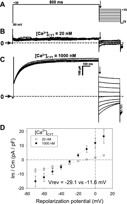
Fig. 3.
Conductance of murine CaCC. A: protocol used to measure Ca2+-dependent Cl− conductance. Pericytes were held at −80 mV and activated by depolarization to +50 mV for 800 ms. Subsequently, cells were repolarized to test potentials between +10 and −70 mV. B and C: current traces obtained with electrode [Ca]CYT of 20 or 1,000 nM. Horizontal arrows and dashed lines indicate zero current level. D: normalized membrane current vs. repolarization potential, measured immediately after decay of capacitance transient (vertical arrows in A–C), yields slope conductance and reversal potential (Vrev) when electrode [Ca]CYT is 20 or 1,000 nM. Vrev shifted toward Nernst potential of Cl− when [Ca]CYT was raised.

