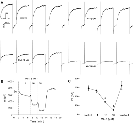
Fig. 6.
Blockade of murine CaCC by the myosin light chain kinase inhibitor ML-7. A: pericytes were held at −80 mV and sequentially depolarized to +70 mV for 1,000 ms at 10-s intervals (inset). Concatenated examples (interpulse intervals omitted) of currents elicited during depolarizations at baseline and during exposure to ML-7 (1, 10, and 50 μM) are shown. B: end-pulse current as a function of time during exposure to ML-7. Inhibition was concentration-dependent and reversible. C: summary of end-pulse current before, during, and after exposure to increasing concentrations of ML-7 (n = 6). *P < 0.05 vs. baseline (control).

