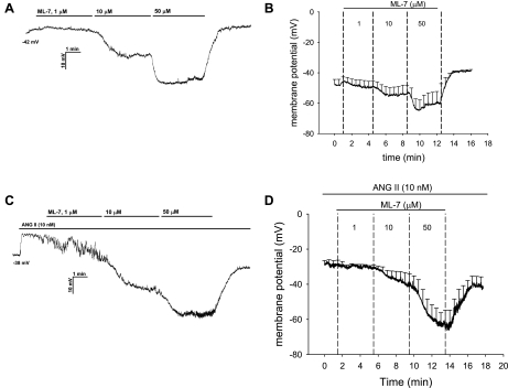
Fig. 7.
Repolarization of ANG II-stimulated murine pericytes by ML-7. A and B: example and summary (n = 5) of nystatin perforated-patch recording of pericyte membrane potential at baseline followed by sequential addition of ML-7 to the extracellular buffer at 1, 10, and 50 μM. C and D: example and summary (n = 6) of membrane potential recording during exposure to ANG II (10 nM) followed by sequential, concomitant addition of ML-7 at 1, 10, and 50 μM. Values are means ± SE; most error bars are suppressed for clarity. ML-7 hyperpolarized resting pericytes and reversibly repolarized ANG II-stimulated cells.

